pleural effusion cat ultrasound
Ultrasound examination of the heart echocardiogram Laboratory tests Treatment Pleural Effusion in Cats The treatment of pleural effusion ultimately will depend upon the underlying cause. In the latter situations therapeutic intervention must be initiated quickly to prevent respiratory arrest.

Cat Of Figure 1 Thoracic Ultrasound Revealed A Mild Hypoechoic Download Scientific Diagram
In contrast to humans veterinary studies have shown questionable sensitivity and specificity for TFASTandVet BLUEtodetectpleuralspaceandlungpathology12.

. To characterize the effusion noting echogenicity of the fluid any loculations solid masses and pleural disease. Ultrasound protocols are highly sensitive and specific for the detection of pleural effusion alveolar interstitialsyndromeandpneumothorax. Lung ultrasound findings including pleural effusion PLEFF number of Blines and subpleural abnormalities were noted.
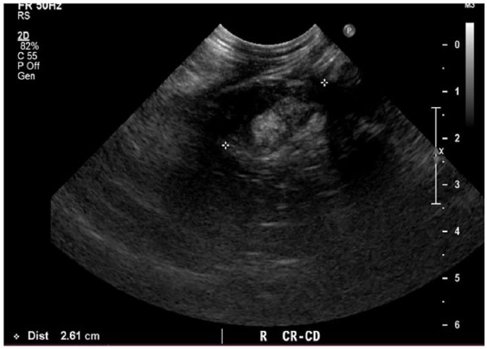
The caudal vena cava cvc is seen extending from the liver L to the heart H. There are a number of characteristic findings on radiographs that will help your veterinarian identify the presence of pleural effusion. TFAST is used for rapid detection of pneumothorax and pleural and pericardial effusion.
Initial treatments may vary depending on the likelihood of the specific diseases based on your pets physical examination and history. Pleural effusion is present in both hemithoraces e. Fluid Scoring System TFASTÒ for the detection of pleural and pericardial effusion pneumothorax and its 4 TFASTÒ echo views and Vet BLUEÒ the veterinary brief lung ultrasound exam a regional pattern-based approach with its B-line Scoring System and its Visual Lung Language.
Abdominal abnormalities identified on ultrasound included abdominal masses lymphadenopathy hepatic venous congestion hepatomegaly splenomegaly renal enlargement small intestinal wall thickening steatitis and pancreatitis. Histopathology and cultures revealed fungal pneumonia and pyothorax caused by Aspergillus fumigatus. The aims of ultrasound guided assessment of pleural effusion are.
Pleural effusion in cats was commonly reported to be associated with neoplasia and its presence usually indicates guarded prognosis and short survival 2932. Patients most commonly present with dyspnea initially on exertion predominantly dry cough and pleuritic chest pain. The therapeutic intervention also provides your first diagnostic test.
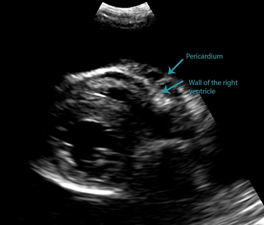
Careful handling and prompt and adequate stabilisation incorporating supplemental oxygen. Cats presenting with pleural effusion are nearly always in respiratory distress ranging from an increased respiratory rate and effort to open mouth breathing. In the moving clip however you can actually see the separation of the right ventricular free wall from the pericardium in a cat.
In some cases ultrasound may also be. Additional findings in patients with pyothorax may include depression anorexia weight loss dehydration muffled heart and lung sounds and pale mucous membranes. There is some fluid in the pericardium but this is normal and only seen during systole.
Malignant pleural deposits or primary lung neoplasms as well as subdiaphragmatic diseases eg. It can pose a diagnostic dilemma to the treating physician because it may be related to disorders of the lung or pleura or to a systemic disorder. Pleural effusion volume mL distance cm x 90 the second Goecke formula measures the distance between the lung base and the mid-diaphragm the subpulmonary height height of the dome of the diaphragm is connected to the lung base the line being perpendicular to ones field of insonation.
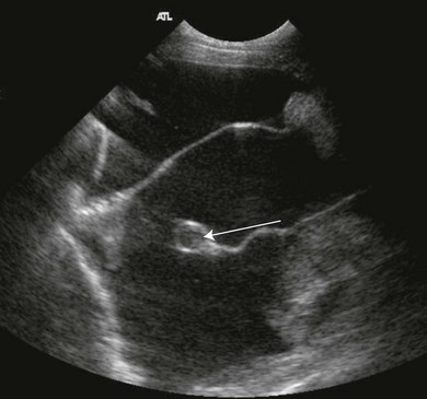
Caudal is to the left of the image. B Longitudinal ultrasound scan of the caudal thorax of a cat with pleural effusion. Refer to the article Pleural effusion volume ultrasound for more information CT CT scanning is excellent at detecting small amounts of fluid and is also often able to identify the underlying intrathoracic causes eg.
Cats with pleural effusion often have severe respiratory compromise at presentation. A sample of pleural fluid obtained by piercing the cats chest cavity with a needle will be sent to the laboratory for analysis. The success rate is low when the effusion is loculated and septated.
Ultrasound-guided pleural effusion drainage by catheter insertion is a safe and effective procedure. It is worth evaluating the sonographic appearance of pleural effusion for predicting neoplastic etiology. The trocar technique is faster and easier.
Blood NTproBNP LUS and FCU evaluating left atrial LA size and presence of pericardial effusion PCEFF were performed in all cats. To mark the optimal site for drainage and perform the procedure if required. X-ray and ultrasound imaging of the chest cavity are also very helpful in analyzing the causative factors.
Bilaterally applied chest tube site and pericardial site views plus diaphragmatico-hepatic view also part of AFAST Vet BLUE. Medical records were evaluated for final diagnosis. To determine and describe the size and site of the effusion.
Abdominal ultrasound repeat thoracic radiography urinalysis with culture and retroviral screening failed. Thoracic ultrasound revealed a pleural effusion and a focal lung mass. Found with right congestive heart failure obstruction to lymphatic drainage by tissue adhesions in pleural space lung lobe torsion neoplasms and abdominal contents herniating.
Each of these 3 ultrasound formats has. Ultrasonography is superior to chest radiography in detecting the presence of pleural effusions and in distinguishing pleural effusions from atelectasis or pleural thickening. In this cat below it is difficult at first glance to be able to state whether this effusion is pleural or pericardial.
Both the trocar and the modified Seldinger techniques can be used. The chest wall may seem incompressible in cats with thoracic effusion. Four standard effusion types recognized in addition to blood.
The transducer is perpendicular to the ribs. TFAST a standardized and validated thoracic point-of-care ultrasound examination includes 5 acoustic windows. Pleural effusion is typically diagnosed by taking radiographs X-rays of the chest.
The type of pleural fluid withdrawn will enable your veterinarian to diagnose the cause of the pleural effusion. This procedure removes excess fluid from the pleural space using a needle which not only relieves pressure on your cats lungs but also provides your veterinarian with pleural fluid samples. A pleural effusion is an excessive accumulation of fluid in the pleural space.
The transducer is perpendicular to the ribs. The cat underwent exploratory thoracotomy and a total left pneumonectomy was performed. If the FAST ultrasound does reveal pleural effusion thoracentesis can be carried out.
Abdominal ultrasounds were performed in 70 cats with pleural effusion and revealed concurrent abdominal effusion in 59 of these cats. Thoracic radiographs usually reveal pleural effusion. Accumulation of fluid in the pleural space.
Compared to chest computed tomography CT scan pleural ultrasound has 93 sensitivity and specificity for detecting pleural effusions.
Spontaneous Cholecystopleural Fistula Leading To Biliothorax And Sepsis In A Cat
Differentiating Pericardial From Pleural Effusion Animal Ultrasound Association
Pleura Veterian Key

Different Types Of Pleural Effusion On Ultrasound Scan A Exudate B Download Scientific Diagram

How To Ultrasound Detection Of Pleural Fluid Case Study Video Youtube

Differentiating Pericardial From Pleural Effusion Animal Ultrasound Association

Search Imv Imaging

Veterinary Echocardiography Newsletter 1 Effusions Animal Ultrasound Association
Pdf Thoracic Ultrasound A Method For The Work Up In Dogs And Cats With Acute Dyspnea Semantic Scholar